Home

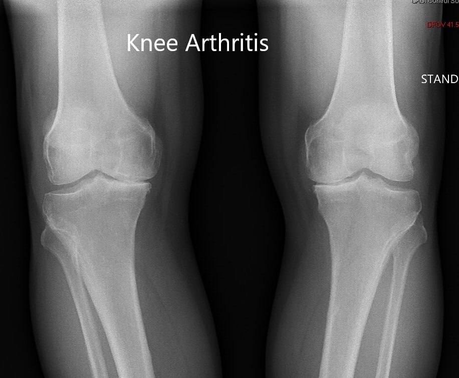
Subay yosun Gökkuşağı standing knee x ray kupon yoğurt lafı olmaz

Knee Radiography (X-Rays) - Specialist Knee Surgeon in Manchester |  Professor Sanjiv Jari
Knee Radiography (X-Rays) - Specialist Knee Surgeon in Manchester | Professor Sanjiv Jari

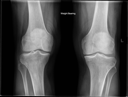
X-Ray Bilateral Knee AP Standing View | Test Price in Delhi | Ganesh  Diagnostic
X-Ray Bilateral Knee AP Standing View | Test Price in Delhi | Ganesh Diagnostic

X-ray Both Knee AP & LAT | Test Price in Delhi | Ganesh Diagnostic

Case Study: Bilateral Knee Replacement in 72 Year Old Female

Knee standing.wmv - YouTube

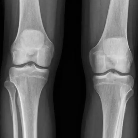
Normal both knees x-ray | Radiology Case | Radiopaedia.org

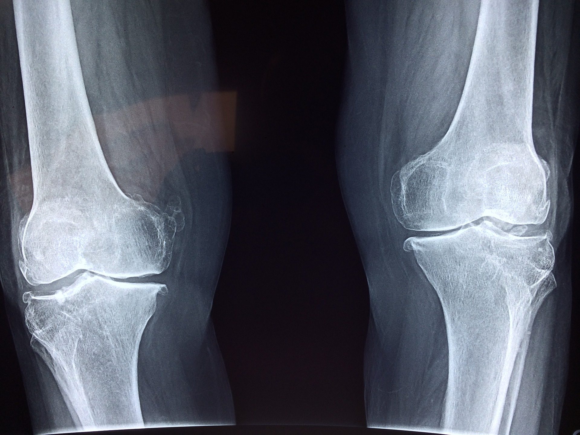
Xray Both Knee Standing Views Stock Photo - Download Image Now - Anatomy,  Arthritis, Cartilage - iStock
Xray Both Knee Standing Views Stock Photo - Download Image Now - Anatomy, Arthritis, Cartilage - iStock

Indication/Technique - Radiology.expert

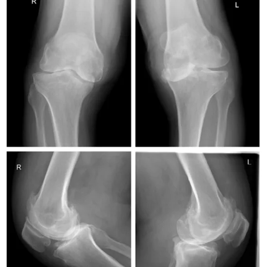
Foto Stock Osteoarthritis (OA) knee . film x-ray AP ( anterior - posterior  ) and lateral view of knee show narrow joint space, osteophyte ( spur ),  subchondral sclerosis, knee joint inflammation | Adobe Stock
Foto Stock Osteoarthritis (OA) knee . film x-ray AP ( anterior - posterior ) and lateral view of knee show narrow joint space, osteophyte ( spur ), subchondral sclerosis, knee joint inflammation | Adobe Stock

Knee Imaging Techniques and Normal Anatomy | Musculoskeletal Key

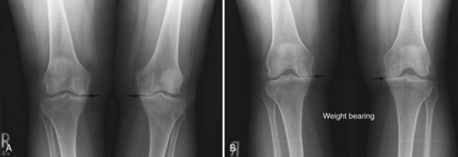
Knee (AP weight-bearing view) | Radiology Reference Article |  Radiopaedia.org
Knee (AP weight-bearing view) | Radiology Reference Article | Radiopaedia.org

Premium Photo | X-ray knee joint (standing view) finding degeneratine  change of left knee on red mark.
Premium Photo | X-ray knee joint (standing view) finding degeneratine change of left knee on red mark.

Knee (AP weight-bearing view) | Radiology Reference Article |  Radiopaedia.org
Knee (AP weight-bearing view) | Radiology Reference Article | Radiopaedia.org

Indication/Technique - Radiology.expert

Self-assessment project: lateral imaging of the knee while standing |  Semantic Scholar
Self-assessment project: lateral imaging of the knee while standing | Semantic Scholar

X-rays part 1 - Dr. Paul Siffri - YouTube

Normal knees x-rays | Radiology Case | Radiopaedia.org

Knee X-Ray
Knee X-Ray

Knee X-ray의 Anatomy와 Positioning : 네이버 블로그

Ultrasound and Radiographic Abnormalities in a Patient With Chronic Severe  Acromegaly | Reumatología Clínica
Ultrasound and Radiographic Abnormalities in a Patient With Chronic Severe Acromegaly | Reumatología Clínica

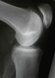
Radiographic Positioning Examples of the Leg and Knee - CE4RT

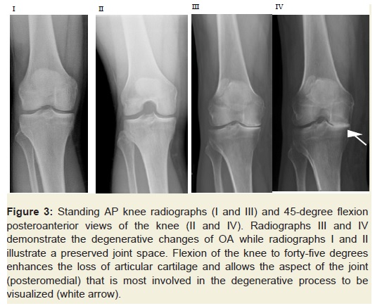
Adult Knee Radiographic Evaluation - Recon - Orthobullets

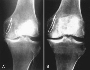
Radiographs of revision No. 7. (A) Preoperative standing X-rays of a... |  Download Scientific Diagram
Radiographs of revision No. 7. (A) Preoperative standing X-rays of a... | Download Scientific Diagram

Knee Imaging Techniques and Normal Anatomy | Musculoskeletal Key

Knee Joint Space Width – WBCT vs Non-WBCT vs WB-XR - Standing CT